
ABEGO coloring. The sheets are in blue, the "B" color (for "beta sheet"). Three loop segments are in red, the "A" color associated with right-handed "alpha" helix. The final loop segment is in green, the "G" color, associated with a left-handed helix, usually involving glycine. The black arrows show the order of the segments.
Foldit uses ABEGO coloring as a guide to how well the backbone angles of a segment correspond to "ideal" angles. (See also Ideal Loops.)
ABEGO coloring appears in several spots. The protein can be shown in ABEGO colors by selecting the "AbegoColor" option available under "Color" on the "View" menu. (This option requires that the "Show advanced GUI" option be checked under "General Options".)
The sequence shown in the Blueprint tool can optionally be shown in ABEGO colors.
The Rama Map tool also shows the "ABEGO" regions as colored areas. When no segments are selected, a generalized ABEGO map is shown. (The generalized map is also shown when multiple segments are selected in the main Foldit window, using the selection interface.) When a single segment is selected, an ABEGO map specific to the amino acid type of that segment is displayed.
When a segment falls within one of the colored regions on the Rama map, it's more likely to be stable and to have a better backbone score.
Each letter in "ABEGO" refers to a "torsion angle class", which is considered significant in the analysis of proteins. Each of the "ABEG" classes is associated with a secondary structure type.
The letters have the following meanings and color assignments in Foldit:
- A - right-handed "alpha helix", red in ABEGO coloring.
- B - right-handed "beta sheet" (or beta strand), blue in ABEGO coloring.
- E - left-handed sheets (very rare), yellow in ABEGO coloring
- G - left-handed helix (rare), green in ABEGO coloring
- O - Cis omega (ω) angle, a special case
The cis case represented by the "O" in ABEGO is different than the other four cases. It doesn't involve the phi and psi angles, so it can't be shown on the Rama map. It doesn't have a color in the ABEGO coloring used in Foldit. In the cis case, the bond between two segments is flipped by 180 degrees from the normal position. The normal bond between two segments (known as the trans case) has an omega angle around 180 degrees. A cis bond has an omega angle around 0 degrees.
The effect of a cis bond is that two sidechains in a row will be on the same side of the protein. A cis bond between two amino acids is most common when proline is the second amino acid.
Examples[]
When ABEGO coloring is used for the main protein, each segment is assigned one of the four colors. This does not mean that the segment falls within the corresponding region on the Rama map. The color intensity of the segment is a guide to how well it's scoring, similar the "hydro/score" color option.
The following examples show cases where the ABEGO color shows problems
Leucine with yellow ABEGO coloring[]

Leucine with yellow coloring associated with left-handed sheets.
In this case, the leucine at segment 6 shows yellow coloring. The Rama map doesn't have a yellow region for leucine, however. In general, glycine is the only amino acid where yellow coloring is good.
Phenylalanine with yellow ABEGO coloring[]
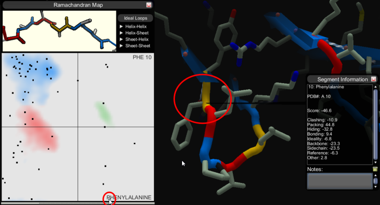
Phenylalanine with yellow coloring associated with left-handed sheets.
The PHE in this example is also well outside its comfort zone.
Valine with red ABEGO coloring[]

Valine with red coloring associated with right-handed helix.
In this example, the valine has red coloring, and the Rama map does have a large red area. This segment is actually slightly outside the red area, however. In theory, this segment might score better if its dot were moved to the right on the Rama map.